OUR SERVICES

CORNEA

The cornea is the clear front surface of the eye. It lies directly in front of the iris and pupil, and it allows light to enter the eye.


Viewed from the front of the eye, the cornea appears slightly wider than it is tall. This is because the sclera (the “white” of the eye) slightly overlaps the top and bottom of the anterior cornea.

The horizontal diameter of the cornea typically measures about 12 millimeters (mm), and the vertical diameter is 11 mm, when viewed from the front. But if viewed from behind, the cornea appears circular, with a uniform diameter of approximately 11.7 mm. This makes the cornea about two-thirds the size of a dime.

The center thickness of the average cornea is about 550 microns, or slightly more than half a millimeter.

The cornea has five layers. From front to back, these layers are:

The corneal epithelium. 

This outer layer of the cornea is five to seven cells thick and measures about 50 microns — making it slightly less than 10 percent of the thickness of the entire cornea. Epithelial cells are constantly being produced and sloughed off in the tear layer of the surface of the eye. The turnover time for the entire corneal epithelium is about one week.

Bowman’s layer. 

This is a very thin (8 to 14 microns) and dense fibrous sheet of connective tissue that forms the transition between the corneal epithelium and the underlying stroma.

The corneal stroma.

This middle layer of the cornea is approximately 500 microns thick, or about 90 percent of the thickness of the overall cornea. It is composed of strands of connective tissue called collagen fibrils. These fibrils are uniform in size and are arranged parallel to the cornea surface in 200 to 300 flat bundles called lamellae that extend across the entire cornea. The regular arrangement and uniform spacing of these lamellae is what enables the cornea to be perfectly clear.

Descemet’s membrane.

This very thin layer separates the stroma from the underlying endothelial layer of the cornea. Descemet’s (pronounced “DESS-eh-mays”) membrane gradually thickens throughout life — it’s about 5 microns thick in children and 15 microns thick in older adults.

The corneal endothelium.

This is the innermost layer of the cornea. The back of the endothelium is bathed in the clear aqueous humor that fills the space between the cornea and the iris and pupil. The corneal endothelium is only a single layer of cells thick and measures about 5 microns. Most of the endothelial cells are hexagonal (six-sided), but some may have five or seven sides. The regular arrangement of these cells is sometimes called the endothelial mosaic.

RETINA

The retina is a thin layer of tissue that lines the back of the eye on the inside. It is located near the optic nerve.The purpose of the retina is to receive light that the lens has focused, convert the light into neural signals, and send these signals on to the brain for visual recognition.


The retina processes light through a layer of photoreceptor cells. These are essentially light-sensitive cells, responsible for detecting qualities such as color and light-intensity. The retina processes the information gathered by the photoreceptor cells and sends this information to the brain via the optic nerve. Basically, the retina processes a picture from the focused light, and the brain is left to decide what the picture is.

Due to the retina’s vital role in vision, damage to it can cause permanent blindness. Conditions such as retinal detachment, where the retina is abnormally detached from its usual position, can prevent the retina from receiving or processing light. This prevents the brain from receiving this information, thus leading to blindness.

UVEA

Uvea is the pigmented middle layer of the eye consisting of the iris and ciliary body together with the choroid.

Eye with uvea. … It has three parts: the iris, which is the colored part of the eye; the choroid layer, which is the layer of blood vessels and connective tissue between the sclera and the retina; and the ciliary body, which secretes the transparent liquid (aqueous humor) into the eye.

uvea (or uveal tract), the middle layer of tissue surrounding the eye that consists of the iris, ciliary body, and choroid. Uveitis can affect people at any age, but onset usually occurs in the third and fourth decades of life.

The posterior portion of the uvea is the choroid, which is sandwiched between the tough outer sclera of the eyeball and the retina in the back of the eye.

Uveitis is swelling of the middle layer of the eye, which is called the uvea. … It’s normally red due to its blood supply from the uvea. Uveitis usually isn’t serious. More severe cases can cause vision loss if not treated early.

CATARACT

The lenses of our eyes are made up of water and protein. This protein helps the light to penetrate through the lens and thus you can see. But with the advancing age these protein gets accumulated in one corner of the lens resulting in the formation of cataract.

Cataracts are mostly common in people over 40-45 years of age, that’s the primary cause of blindness. One of the common symptoms of cataract is blurred vision and it usually occupies a smaller area of the lens in it’s initial stage.

Afterwards it gradually acquires the whole portion of the lens. The few causes that are responsible developing cataract include extensive smoking, uv rays of the sun, obesity and diabetes. Balanced diet, foods enriched with vitamin C and omega-c fatty acids can decrease the chances of cataract formation.

Eye surgeons would remove the cataract placing the artificial lens that would help the patient to regain the lost vision.

Stages of Cataract

Types Of Cataract

cataract-Types

GLAUCOMA

Cupping of optic disc, Raised intraocular pressure, peripheral loss of vision. Glaucoma can be divided into two types: open-angle glaucoma and closed-angle glaucoma. It damages the vision by increasing the pressure of fluid inside the eye. It is also responsible for the damage of retinal ganglion cells.

People of all ages can become a victim and thus one should be careful if facing certain symptoms such as hazy vision, severe headache, nausea or vomiting etc.

Glaucoma when goes to an advanced stage leads to permanent blindness but if detected at an early stage it can be treated using the advanced medical features like Humphery Field Analyser (Perimeter), Applanation tonometer, Rebound tonometer, Pachymetry, Gonioscopy, Optic disc photography, Retinal nerve fibre layers analyser 3D OCT.

PEDIATRIC (CHILD) OPHTHALMOLOGY

Child Eye diseases are unpredictable. Every child ought to go for a general eye check up at the age of 3 that would even help the parents to protect their kid from the unexpected eye problems.

When looking at things are the symptoms of visual disorder in children. Eye problems in children do not only include visual screenings but other problems such as color vision problems, etc.

It’s Parents responsibility to maintain a good vision for your kids that would help you to explore the amazing world with all its colors to your children.

NEURO OPHTHALMOLOGY

Neuro-ophthalmology is the ophthalmic sub specialty that deals with eye diseases and issues caused by different neurological conditions including various optic nerve disorders. This highly specialized and challenging sub specialty is a holistic combination of different primary specialties like Neurology, Neuro-surgery and Radiology with ophthalmology its parent specialty. The Department of Neuro-ophthalmology at V.T. Eye Hospital was started by Dr V. Ramesh, Distinguished Senior Consultant,

The Neuro-ophthalmology department treats a large number of patients with a wide range of symptoms including vision loss, double vision, drooping of eyes, visual field defects, headache, eyelid and facial disorders every day. It also evaluates and treats patients with ophthalmic manifestations of various neurological diseases.

Common pathology referred to a neuro-ophthalmologist includes afferent visual system disorders (e.g. optic neuritis, optic neuropathy, papilledema, brain tumors or strokes) and efferent visual system disorders (e.g. anisocoria, diplopia, ophthalmoplegia, ptosis, nystagmus, blepharospasm, seizures of the eye or eye muscles, and hemifacial spasm).

TRAUMA EYE CARE

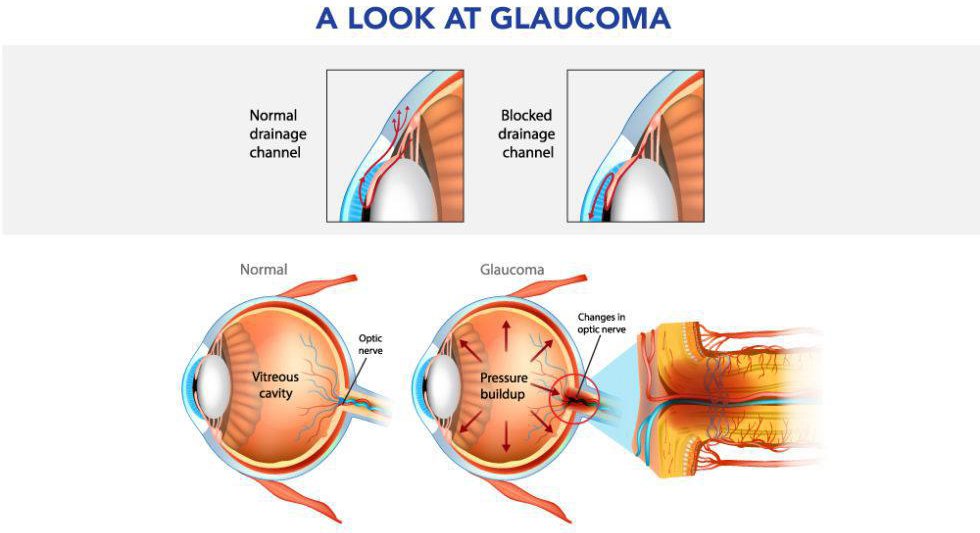
Eye trauma refers to damage caused by a direct blow to the eye. The trauma may affect not only the eye, but the surrounding area, including adjacent tissue and bone structure. There are many different forms of trauma, varying in severity from minor injury to medical emergencies.

Damage to any part of the eye, optic nerve, or any area of the brain related to vision can potentially lead to blindness. One major cause of blindness can be eye injuries, whether physical or chemical. Eye injuries can range from getting a benign and removable substance in the eye to permanent vision loss.

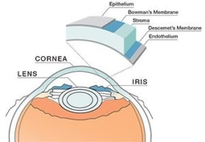
Scratched Eye (Corneal Abrasion)

Common causes of abrasions to the eye’s surface (corneal abrasions) are getting poked in the eye or rubbing the eye when a foreign body is present, such as dust or sand. Corneal abrasions are very uncomfortable and cause eye redness and severe sensitivity to light.

Penetrating Or Foreign Objects In The Eye

If a foreign object such as metal or a fish hook penetrates your eye, visit the emergency room/urgent care center right away. You could cause even more injury to your eye if you attempt to remove the object yourself or if you rub your eye.

If possible, try loosely taping a paper cup or eye shield over your eye for protection; then seek help.

Your eye also may have corneal foreign bodies that are small, sharp pieces of a substance (usually metal) that have become embedded in the eye’s surface (cornea), but have not penetrated into the interior of the eye.

Metal foreign bodies can quickly form a rust ring and a significant scar. Your eye doctor should remove these foreign bodies as soon as possible.

Caustic Foreign Substance In The Eye (Chemical Burn)

Getting unexpectedly splashed or sprayed in the eye by substances other than clean, harmless water can be scary. Some substances burn or sting but are fairly harmless in the long run, while others can cause serious injury. The basic makeup of the chemical involved can make a lot of difference, such as:

Acid :

As a general rule, acids can cause considerable redness and burning but can be washed out fairly easily.

Alkali :

Substances or chemicals that are basic (alkali) are much more serious but may not seem so because they don’t cause as much immediate eye pain or redness as acids. Some examples of alkali substances are oven cleaners, toilet bowl cleaners and even chalk dust.

Chemical exposures and burns are usually caused by a splash of liquid getting in your eye. But they can be caused in other ways as well, such as by rubbing your eyes and transferring a chemical from your hands to your eyes or by getting sprayed in the eye by hair spray or other aerosols.

If you’re splashed in the eye, put your head under a steady stream of barely warm tap water for about 15 minutes. Just let it run into your eye and down your face.

Then call your eye doctor or an emergency room/urgent care center to see what is recommended for your eye injury. Tell the person on the phone exactly what kind of substance got into your eye and what you’ve done about it so far.

If you know your eye is at risk because it’s extraordinarily red or blurry, then just go immediately to your eye doctor or an emergency room or urgent care center after you’ve rinsed it with water. You can put a cool, moist compress or an ice pack on your eye, but don’t rub it.

Depending on the substance, the effects of chemical exposures causing eye injuries can range from minor irritation and red eyes to serious eye damage and even blindness.

Eye Swelling

Eye swelling and puffy, swollen eyelids can result from being struck in the eye such as from a baseball moving at a high speed.

The best immediate treatment for this type of eye injury is an ice pack.

You may have a simple black eye (bruising around the eye), but you should see an eye doctor to make sure there’s no internal damage.

Subconjunctival Hemorrhages (Eye Bleeding)

This eye injury usually looks worse than it really is. A subconjunctival hemorrhage involves leakage of blood from one or more breaks in a blood vessel that lies between the white of the eye (sclera) and its clear covering (conjunctiva).

Subconjunctival hemorrhages are quite common and can occur from even minor injury to the eye. They may be limited to a small sector of the eye, or they can extend over the entire eye, making the white sclera appear bright red.

A subconjunctival hemorrhage is painless and does not cause temporary or permanent vision loss. No treatment is required. Over the course of several weeks, the blood will clear and the eye will return to a normal appearance.

Traumatic Iritis

Traumatic iritis is inflammation of the colored part of the eye that surrounds the pupil (iris) and occurs after an eye injury. Traumatic iritis can be caused by a poke in the eye or a blow to the eye from a blunt object, such as a ball or a hand.

Traumatic iritis usually requires treatment. Even with medical treatment, there is a risk of permanent decreased vision.

Hyphemas And Orbital Blowout Fractures

A hyphema (high-FEE-mah) is bleeding in the anterior chamber of the eye, the space between the cornea and the iris. Orbital blowout fractures are cracks or breaks in the facial bones surrounding the eye.

Hyphemas and blowout fractures are serious eye injuries and medical emergencies. They are caused by significant blunt force trauma to the eye and face, such as getting hit by a bat, baseball, hockey stick or puck, or getting kicked in the face.

Steps To Take In Case Of Eye Injury

  1. If you have any eye injury, contact your eye care practitioner immediately for advice.
  2. Most eye doctors have emergency contact numbers for injuries that occur after normal business hours or on weekends.
  3. In certain extreme situations such as a penetrating eye injury or an eye knocked out of the socket, it may be better to get to the hospital immediately without taking the time to try calling anyone.
  4. Once you are in the care of a doctor, be sure to mention if you wear contact lenses so you can be advised whether to leave them in or remove them.
  5. Depending on the type of eye injury, the doctor may want you to flush your eye with water or saline solution. In more serious situations, you may need surgery.
  6. Treat all eye injuries as potential emergencies, and never hesitate to contact or see an eye doctor immediately. Don’t take risks with your eyesight. Remember, you have only one pair of eyes.

SURGERY

Operation Theater

The operating room is equipped with sophisticated medical equipment to deal with every situation, from the simple to the most intricate procedures.

Pain care center experts in pre & post operative pain management services.

Operation

Eye surgery also known as ocular surgery is surgery performed on the eye or its adnexa, typically by an ophthalmologist. The eye is a very fragile organ and requires extreme care before during and after a surgical procedure to minimise or prevent further damage.

An expert eye surgeon is responsible for selecting the appropriate surgical procedure for the patient, and for taking the necessary safety precautions.

  • Preparation and precautions
  • Cataract laser surgery
  • Glaucoma surgery
  • Canaloplasty
  • Refractive surgery
  • Corneal surgery
  • Vitreoretinal surgery
  • Eye muscle surgery
  • Eyelid surgery
  • Orbital surgery
  • Surgery involving the lacrimal apparatus
  • Eye removal

Other surgery

  1. Ciliary body in an attempt to control glaucoma
  2. Since highly safer surgeries for glaucoma
  3. Including lasers
  4. Nonpenetrating surgery
  5. Guarded filtration surgery
  6. Seton valve implants have been invented

OPTICAL & PHARMACY

Optical

Well stocked branded Frames, Lenses in different models and Eye Ware Accessories, Sunglass, Therapeutic and Cosmetic Contact Lenses with the help of professional Opticians

Pharmacy

Well-stocked pharmacy with an entire range of surgical and medical products to meet every need of the patient.

Contact Lens